What is an OCT scan?

An optical coherence tomography scan (commonly referred to as an OCT scan) helps us to view the health of your eyes in greater detail, by allowing us to see what’s going on beneath the surface of the eye.

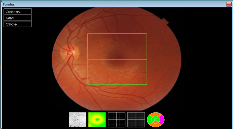
Imagine it like a cake – we can see the top of the cake and the icing using the 2D digital retinal photography (fundus camera), but the 3D image produced from an OCT scan slices the cake in half and turns it on its side so we can see all the layers inside.

Our opticians can then map out and measure the thickness of these layers to get an even clearer idea of your eye health.

OCT scans can help detect sight-threatening eye conditions earlier. In fact, glaucoma can be detected up to four years earlier.

OCT is separate to an eye test. An eye test checks your eye health as well as how well you can see. Part of that often involves taking an image of the back of the eye (digital retinal photography), but an OCT scan takes this a step further, allowing your optician to look even deeper into your eyes and the structures within it.

Essentially, an OCT scan gives your optician a clearer idea of your eye health when testing your eyes.

Why do I need an OCT scan?

OCT scans are recommended for people aged 25 or over, who want to know more about their eye health, or those who have diabetes, glaucoma, or have family history of eye disease.

Even if your vision and eye health are perfectly fine, we still recommend an OCT scan with every eye test. It’s really useful for your optician to have a baseline image on file, so they can monitor any changes over time. It’s the same as when we take an image of the back of your eye with digital retinal photography.

So the next time you come in for an eye test, your optician can compare your images from your last visit as well as comparing measurements to averages and might spot even the tiniest change in the eye’s structures. This could help indicate the early signs of an eye health condition like glaucoma, diabetic retinopathy and macular degeneration before you have any symptoms.

This means that conditions like these can be managed before they get worse and can help prevent potential sight loss. That’s a pretty good result for just a quick scan.
How much does an OCT scan cost?
Because OCT scans are separate to your eye test, an additional charge may apply so please speak to us about this when booking your appointment.
Does it take longer than a regular eye test?
An OCT scan takes a matter of seconds and your optician will go through the results with you during your eye examination.

What is an OCT scan used for and what conditions can it help to detect?

OCT scanning is great at confirming that your eyes are healthy and can be repeated over time for comparison. This makes it particularly useful for detecting potentially sight threatening conditions that generally don’t have any symptoms until they start to have an impact on your vision.

Many of these conditions form at the very back of the eye, and OCT allows your optician to identify subtle changes over time, such as changes in the vitreous (the jelly-like substance that fills the eye), retina (the light-sensitive layer at the back of the eye), macula (an area at the centre of the retina responsible for our central vision) and the optic nerve (transmits light impulses to the brain to produce the images we see).

These include:

  • Glaucoma
  • Diabetic retinopathy
  • Detached retina
  • Age-related macular degeneration
  • Macular hole

In fact, an OCT scan can help to detect glaucoma up to four years earlier than traditional methods.

How does optical coherence tomography work?
Taking just a few seconds, an OCT scan uses light to take over 1,000 images of the back of your eye and beyond, looking right back to the optic nerve.

A layered image is created that gives us an incredibly accurate picture of your eye and its structures, allowing us to check your eye health. The images will then be stored so we can note changes over time.
I have a diabetic check annually; do I still need an OCT scan?
Yes – while both the diabetic screening check and OCT involve taking images of the back of the eye, there are significant differences.

Diabetic checks involve a fundus picture – this is an image of the surface of the back of the eye (the retina) also known as digital retinal photography. OCT images allow us to look at the many layers beneath the surface of the retina, which helps us to spot changes to eye health earlier than just looking at the surface.

OCT scans also help in the detection of a range of other eye problems, not just those that are linked to diabetes.